|
Structural MRI & EEG |
|
 J. S. * 74 ‚Helene’
comparison to other cases |
| Structural MRI: |
Brain routine MRI without contrast was recorded on 4-05-04, at Hennepin County Medical Center (MN). Findings: No mass-effect or midline shift, but encephalomalacia in the left frontal lobe and mild T2 hyperintensity within the left middle frontal gyri; ventricles not enlarged. No evidence of hemosiderin or chronic blood product deposition. |
|
| EEG: |
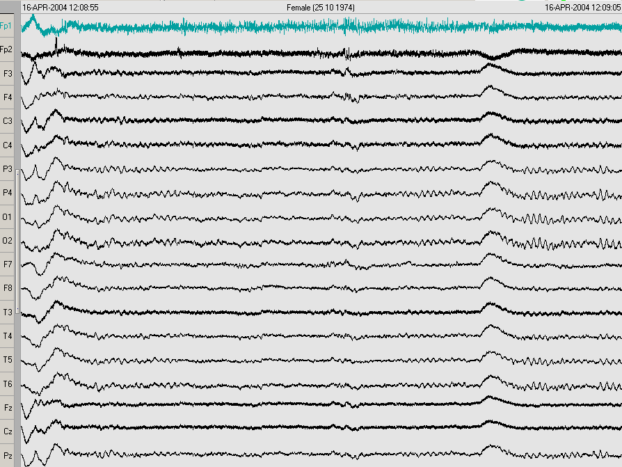
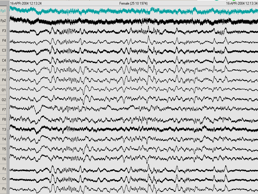
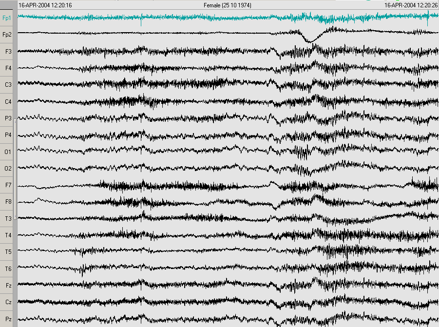
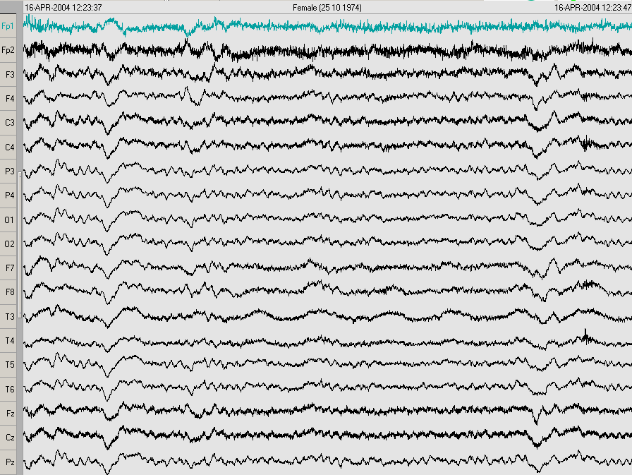
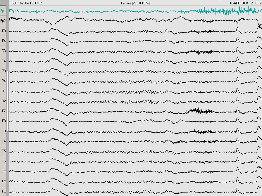
Awake and sleep 20-channel EEG recorded on 4-16-04, at Hennepin County Medical Center (MN), including 3 min hyperventilation, and photostimulation. Well-developed posterior rhythm of 10 Hz (a). Diffuse fast activity, with higher amplitude in the left anterior quadrant. Normal descent into stage 2 sleep, with symmetrical vertex waves and spindles. No epileptiform features. |
|

|
awake, eyes open |
awake, eyes open, high frequency |
awake, eyes open, after 3 min hyperventilation |
awake, eyes closed / open, after photic stimulation |
|
|
|